Anatomia

The body as a machine

Le proporzioni del corpo umano

Leonardo studied people the way he studied his machines: by taking them apart. Around 1490 he measured the body against a Roman architect’s rules. Twenty years later, in the winter of 1510–11, working with the Pavia anatomist Marcantonio della Torre, he may have dissected as many as twenty bodies, drawing what he found more clearly than anyone had before.

He meant to publish a treatise. Then war came to Milan, della Torre died of the plague in 1511, and the drawings went unseen for centuries. About 150 of his anatomical sheets survive, most of them now in the Royal Collection at Windsor.

scroll, and the folios draw themselves ↓

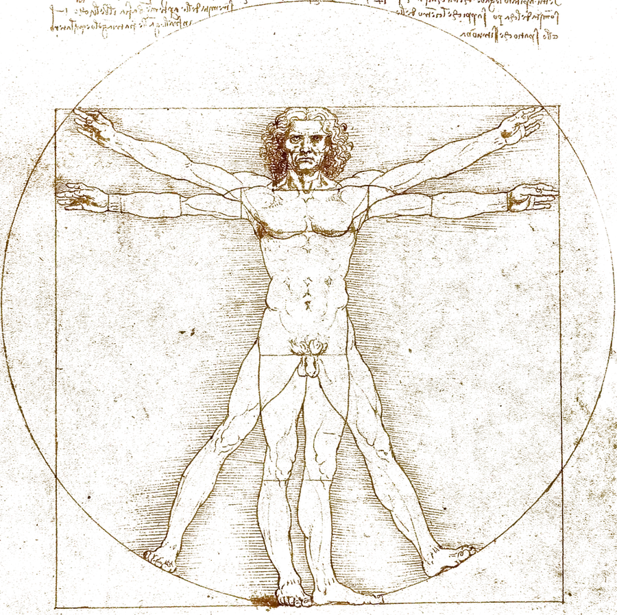
Leonardo’s Vitruvian Man: a man in two poses inside a circle and a square.

Folio I · c. 1490

The Vitruvian Man

A Roman architect, Vitruvius, wrote that a well-made man fits both a circle and a square. Others had tried to draw it and failed. Leonardo made it work by drawing the man twice, in two poses, with a different centre for each shape.

Source · Leonardo’s own lines, traced from the Vitruvian Man (Gallerie dell’Accademia, Venice) and redrawn stroke by stroke; the red measurements are ours.

Was he right?

An ideal, not a man

These are the ideal proportions Vitruvius described, refined by Leonardo’s own measuring, not a rule every body follows. Real people vary.

What he got right
The geometry. Earlier drawings centred both shapes on the navel and could not make it fit; he centred the circle on the navel and the square on the genitals.
le braccia apertel’altezzaun ottavol’ombelicothe navelil cerchiothe circleil centro del quadratocentre of the square

Folio II · 1489

The Skull

Among his earliest anatomy, two decades before his great dissections: the skull from the front, its left half cut away down the middle to show the hollows behind the face, the eye and the cheek. On a sheet from the same set he placed a skull where three lines at right angles meet, a way of locating what lies inside.

Source · Leonardo’s own lines, traced from the skull sectioned (Royal Collection, RCIN 919058, verso) and redrawn stroke by stroke; the red notes are ours.

Was he right?

A first step

These sheets come long before his real dissections of 1510–11. The skull is where he started, treating the head as a space to be measured like a building.

What he got right
Mapping the inside of the head with a grid. A sheet from the same set opens with the note “On the 2nd day of April 1489”.
la metà segatathe sawn halfocchioeye socketdentiteethle cavitàthe hollows

Folio III · c. 1510–11

The Spine

The column from the side and from the front: seven vertebrae of the neck, twelve of the back, five of the loins, then the fused sacrum. His own letters down the side mark where each region ends.

Source · Leonardo’s own lines, traced from the vertebral column (Royal Collection, RCIN 919007, verso) and redrawn stroke by stroke; the red brackets are ours.

Was he right?

Yes

The Royal Collection calls it the first accurate depiction of the spine in history: its sections and curvatures are all correct.

What he got right
The S-shaped curve and the count of vertebrae. His one slip, in the upper back: twelve vertebrae, but thirteen spines.
sette del colloseven of the neckdodici del dorsotwelve of the backcinque dei lombifive of the loinsl’osso sacrothe sacrumuna spina di troppoone spine too many

Folio IV · c. 1512–13

The Heart’s Valve

Where blood leaves the heart, three cusps open and close. Leonardo watched flow through a model of the valve and saw the blood curl into eddies in the pouches behind the cusps, and reasoned that those eddies help the valve shut.

Source · Leonardo’s own lines, traced from his studies of the aortic valve (Royal Collection, RCIN 919082) and redrawn stroke by stroke; the turning eddies are ours, set on his.

Was he right?

Yes

Five centuries later, imaging of living hearts showed the eddies in the aortic root where he had drawn them.

What he got right
That swirling blood in the sinuses plays a part in closing the valve.
Tested since
In 2014 an Oxford team used 4D MRI on living hearts: “Flow vortices in the aortic root: in vivo 4D-MRI confirms predictions of Leonardo da Vinci”, European Heart Journal. Bissell et al., 2014
la valvolathe valvei vorticithe eddiesil senothe sinusl’aortathe aorta

Folio V · c. 1510–11

The Shoulder and Arm

The same shoulder four times, turned a step further each time from the front towards the back. With a second sheet the sequence runs to eight views, and in the corner he drew a star of eight points to plan them: a way of showing the body in the round that no single drawing could.

Source · Leonardo’s own lines, traced from the muscles of the shoulder, arm and neck (Royal Collection, RCIN 919008, verso) and redrawn stroke by stroke; the red marks are ours.

Was he right?

Yes, nearly all

The Royal Collection notes that every surface muscle of the upper arm and shoulder on these sheets can be identified. His one quirk: he divided the deltoid over the shoulder into separate pieces of his own.

What he got right
The muscles themselves, and the method: turning one subject through eight views to show it whole.
il deltoidethe deltoidil deltoide divisothe deltoid, dividedgli otto aspettithe eight views

Misuralo · Measure the real one

His sheet, under the ruler

This is Leonardo’s own drawing, not ours. Choose a rule and it is drawn over his sheet, so you can check it against his figure.

Measure from the soles of the feet to the top of the head, then across the outstretched arms: the two are the same, so a man fits a square.

Vitruvius, De architectura, Book III

Leonardo da Vinci, the Vitruvian Man: a man in two overlapping poses inside a circle and a square, with his notes above and below.
The Vitruvian Man, c. 1490. Pen and ink on paper, 34.4 × 25.5 cm.
Gallerie dell’Accademia, Venice · public domain · view on Wikimedia CommonsThe red lines are ours, drawn over his.

Altri fogli · More of his sheets

The fetus in the womb, c. 1511 (RCIN 919102).
Royal Collection Trust, Windsor · public domain · Commons

Il feto · c. 1511

The child in the womb

A child curled inside the opened womb, with smaller sketches and notes on reproduction around it.

What he got right
He is credited as the first to draw the fetus in its correct position in the womb.
What he got wrong
The placenta is drawn as many separate pieces, the way he had seen it in a cow. He never found that the human placenta is a single disc.

Sources: Royal Collection, RCIN 919102 · Embryo Project, Arizona State University

Fonti · Sources

The drawings in the folios are Leonardo’s own lines: his ink was lifted from the public-domain photographs of his sheets and traced into pen paths, so the page can draw it again in the order a pen would move. The red marks, brackets and moving eddies laid over them are ours, and each folio’s toggle shows the whole sheet. The Italian captions are ours, written in his manner, not his words. The photographs of his sheets come from the institutions that hold them, shared as public domain on Wikimedia Commons. The red measurements are ours. The skeleton, skull, spine, heart, shoulder and arm you can take apart in three dimensions are not his: they are a modern anatomical model, BodyParts3D, simplified for the web and shared under the same licence. The glass valve in the heart’s room is ours.

Sources: Royal Collection, RCIN 919058 · Royal Collection, RCIN 919057 · Royal Collection, RCIN 919059 · Royal Collection, RCIN 919008 · Royal Collection, RCIN 919007 · Royal Collection, the aortic valve · Bissell et al., 2014 · Royal Collection, RCIN 919102 · Embryo Project, Arizona State University · Nature: Leonardo’s anatomy · Royal Collection: Leonardo’s study of anatomy · Wikipedia: Vitruvian Man · BodyParts3D, © The Database Center for Life Science, CC Attribution-Share Alike 2.1 Japan (3D skeleton, skull, spine, heart, shoulder and arm; meshes from the Kevin-Mattheus-Moerman/BodyParts3D mirror, simplified for the web)